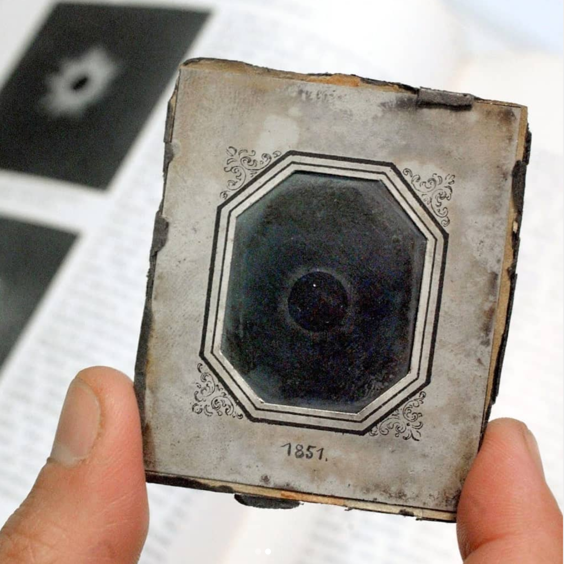
The first image of a total solar eclipse was taken in 1851 (169 years ago). Daguerreotypy was used to capture the moment – a process that involves a copper plate with silver on it that is made light sensitive, exposed to light in camera, developed, then preserved. All sorts of chemicals were used and the camera back then was a simple box with a hole (camera obscura).
Relating this to biology: while microscopes seem to have developed separately, cameras and microscopes converged right before1900. Until then, scientists drew their images, sometimes tracing it (with a camera lucida). Initially called photomicrography, attempts of obtaining photographs through a microscope date back to 1802. Beginning in the 1840’s though, Daguerreotypes (described above) became used for imaging through the microscope. These images seem to have been more stable as earlier photographs did not survive – ironically only drawn copies remain. From the 1850’s, the imaging technique improved in parallel with camera development. Yet, even in the 1950’s (a century later), the number of microscopic photographs were still low (lithography, where initial steps include drawing, was a popular alternative). However, by the turn of the century, photographs dominated published science articles and have ever since.
Solar eclipse image by Julius Berkowski, 1851 (photograph of it from Time)
Picromicrography summary from:
Bradbury, S. (1994). Recording the image – past and present. Quekett Journal of Microscopy. 37: 281–295.
Originally posted on Instagram March 25, 2020

No Responses