I want to mention a couple key techniques in neuroscience:
Calcium imaging
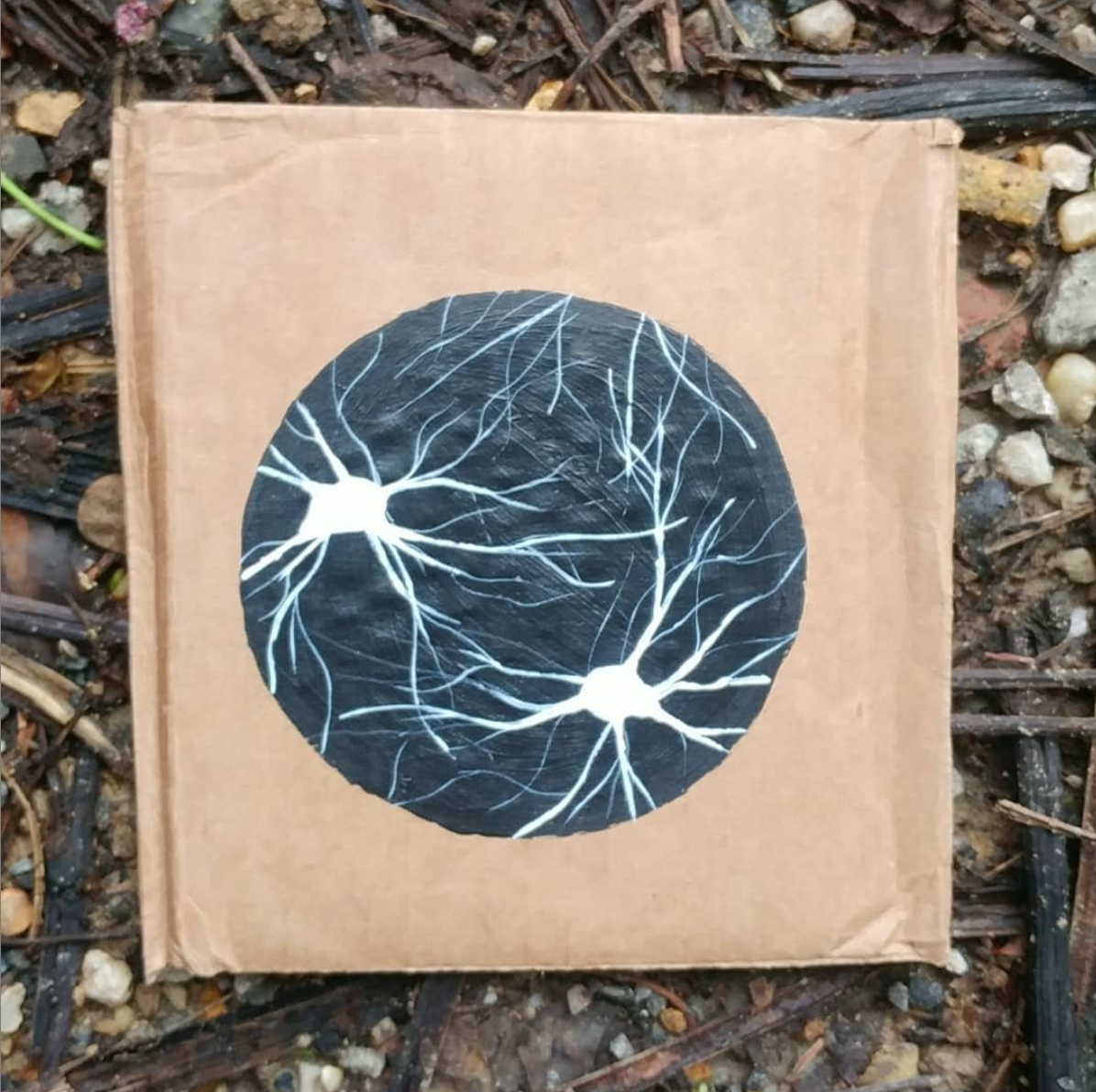
TLDR; you can visualize stimulated neurons by putting in a protein that “glow” with activity!
Neurons “fire.” This is biology jargon for cells having a specific type of activity, like when neurons communicating between each other. Communication between neurons can happen in a variety of different forms, like passing chemicals (neurotransmitters) among themselves. That’s cool and all, but these different forms of communication usually needs the cell to “fire.” Firing refers to, simply put, when the voltage of a cell changes and there is an influx of calcium. If there are proteins that detect calcium presence within the cell, we can then visualize when the cell is active. This protein, called GCaMP, becomes fluorescent green as a cell fires. Therefore, when a cell is active, you can see what looks like slow fireworks under the microscope.
There are slightly modified versions of GCaMP, like jRGECO and RCaMP (both are red fluorescent calcium indicators). There are also voltage sensors, which will more directly measure neuronal activity, but GCaMP is most developed with most widely used. Knowing when cells are active are key to understanding how behaviors arise.

Optogenetics
TLDR; light-sensitive proteins that make neurons active with light!
This is a really popular technique to understand what certain cells do in behavior and is often used in worms (C. elegans ), mice, and fruit flies (drosophila). Cells fire when communicating between themselves and that this activity is characterized by a voltage change and an increase in calcium. The voltage change is actually caused by a sudden rush of positively charged ions (sodium) into the cell. The idea behind optogenetics is to have proteins that allow sodium to enter the cell with a flash of a (blue) light. This light-sensitive protein is called rhodopsin and it was originally found in algae.
There are different types of proteins available now to activate (make cells fire) or inhibit (make cells less likely to fire), with different speeds, and with different colors. Some of them have really fun names like, Chrimson for a red-light sensitive proteins and ChETA (pronounced “cheetah”) for super fast proteins
Researchers all over the world are using optogenetic techniques to deconstruct behaviors. For example, activating some cells in the hypothalamus makes mice start eating, but making another cell type in the same brain area makes them stop eating. Understanding the what regulates when an animal eats might help understand our current obesity epidemic. Maybe learning about why people gain weight is a bit silly, but obesity is obviously a problem (over a third of American adults are obese) and its associated with all sorts of health problems, which costs a lot of money.

Summary
Some researchers are able to combining calcium imaging with optogenetics, which can be incredibly powerful to see how neurons are functionally connected to each other. These are cutting edge (and expensive) techniques, but there are many ways of learning when a neuron is active without imaging individual cells. For instance, there is fiber photometry (this takes the average fluorescence of the area with GCaMP expressing neurons, removing any single-cell information) and electrophysiology (this measures the electrical signal throughout an area). You can also stimulate cells with electricity, but the electricity-based techniques won’t let you pick out which cell types to target in a given area. Observing and manipulating specific populations of neurons is largely where neuroscience is headed since there is already decades of research studying the effects of zapping and cutting different large brain areas.
Originally posted on Instagram June 11-5, 2018 (3 posts)
No Responses